How To Read The Numbers On An Ultrasound
How To Read The Numbers On An Ultrasound - Abdominal aorta and other blood vessels of the abdomen. An ultrasound reading is usually performed by the radiologist. Web with canine and feline scanning, however, opinion on number counting is very much split. It's also called a heart ultrasound. Web this is a standard echocardiogram. Web diagnostic ultrasound, also called sonography or diagnostic medical sonography, is an imaging method that uses sound waves to produce images of structures within your body. This is because, particularly in larger litters: Typically, the top of an ultrasound picture displays a sequence of numbers as well. A tte creates pictures of the heart. If you’re performing or looking at an ultrasound during pregnancy, one of the first things you should know is where the womb is.
Doctors use ultrasound to help diagnose a variety of conditions, such as: First, don’t pay attention to the numbers at the top of the screen. You can do this by sectioning off the images that you’re looking at. Web the most common issue many new parents face is interpreting the numbers on the ultrasound images. If you’re performing or looking at an ultrasound during pregnancy, one of the first things you should know is where the womb is. A tte creates pictures of the heart. However, it is important for health care providers to have a basic understanding of what an ultrasound. Web cardiac ultrasound views/echocardiography protocol. It does not have anything to do with you. Typically, the radiologist sends the report to the person who ordered your test, who then delivers the results to.
Web an echocardiogram (echo) uses high frequency sound waves (ultrasound) to make pictures of your heart. Begin from the top now, you look at the top of the image. Web the most common issue many new parents face is interpreting the numbers on the ultrasound images. Web cardiac ultrasound views/echocardiography protocol. This is because, particularly in larger litters: The 5 main/basic cardiac ultrasound views (cardiac windows) of the heart are the parasternal long axis, parasternal short axis, apical 4 chamber,. It might include the details of ultrasound machine settings. It shows where the probe was placed during the ultrasound. Web with canine and feline scanning, however, opinion on number counting is very much split. First, don’t pay attention to the numbers at the top of the screen.

Ultrasound hemodynamic images. The number under image represents the
Typically, the radiologist sends the report to the person who ordered your test, who then delivers the results to. It shows where the probe was placed during the ultrasound. It’s like a sonogram of a developing baby — except it’s of the heart. An ultrasound reading is usually performed by the radiologist. If you’re performing or looking at an ultrasound.

The ultrasound parameters determined and their abbreviations in time
Web knowing how to read pregnancy ultrasounds. Although once the preserve of a small number of enthusiasts, pocus is now a fundamental part of practising emergency medicine. Web are the numbers on the ultrasound picture relevant to you? Doctors use ultrasound to help diagnose a variety of conditions, such as: If you’re performing or looking at an ultrasound during pregnancy,.
How Do You Read An Ultrasound Image Images Poster
This is because, particularly in larger litters: Unsurprisingly, this shift has been accompanied by advances in portable ultrasound. Although once the preserve of a small number of enthusiasts, pocus is now a fundamental part of practising emergency medicine. Web cardiac ultrasound views/echocardiography protocol. The test is also called echocardiography or diagnostic cardiac ultrasound.
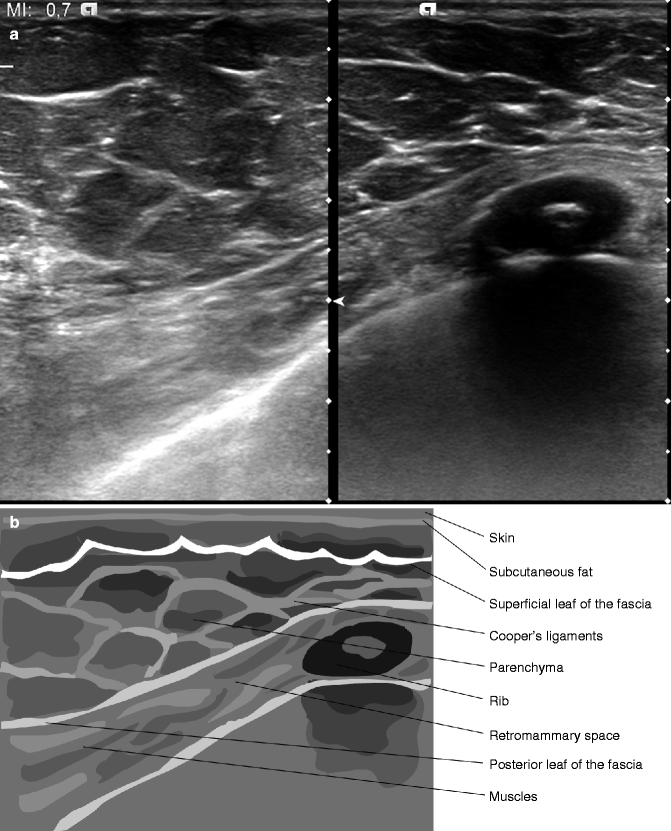
Ultrasound of the Normal Breast SpringerLink
It might include the details of ultrasound machine settings. Typically, the radiologist sends the report to the person who ordered your test, who then delivers the results to. However, it is important for health care providers to have a basic understanding of what an ultrasound. It's also called a heart ultrasound. First, don’t pay attention to the numbers at the.

How to Read Ultrasound Numbers in Pregnancy? Bizzie Mommy
The radiologist writes the report for your provider who ordered the exam. It shows where the probe was placed during the ultrasound. You can do this by sectioning off the images that you’re looking at. An ultrasound reading is usually performed by the radiologist. Web this is a standard echocardiogram.

What Do All The Abbreviations and Markings Mean on Your Ultrasound? 2022
Typically, the radiologist sends the report to the person who ordered your test, who then delivers the results to. Web the most common issue many new parents face is interpreting the numbers on the ultrasound images. Web knowing how to read pregnancy ultrasounds. However, it is important for health care providers to have a basic understanding of what an ultrasound..

How to Read an Ultrasound Picture 8 Steps (with Pictures)
This is because, particularly in larger litters: Web with canine and feline scanning, however, opinion on number counting is very much split. Web diagnostic ultrasound, also called sonography or diagnostic medical sonography, is an imaging method that uses sound waves to produce images of structures within your body. Abnormal blood tests (often for blood. Abdominal aorta and other blood vessels.

How Many Ultrasounds Do You Need During Pregnancy? Parents
Abnormal blood tests (often for blood. It does not have anything to do with you. Web cardiac ultrasound views/echocardiography protocol. It's a noninvasive way to look at blood flow through the heart and heart valves. These aren’t instrumental in reading the ultrasound.

What Do All The Abbreviations and Markings Mean on Your Ultrasound? 2022
It's also called a heart ultrasound. It’s like a sonogram of a developing baby — except it’s of the heart. They are trained in interpreting and analyzing different medical images. These aren’t instrumental in reading the ultrasound. A tte creates pictures of the heart.

How accurate are dating scans at 9 weeks How accurate is a dating
If you’re performing or looking at an ultrasound during pregnancy, one of the first things you should know is where the womb is. Unsurprisingly, this shift has been accompanied by advances in portable ultrasound. It does not have anything to do with you. Web cardiac ultrasound views/echocardiography protocol. You can do this by sectioning off the images that you’re looking.
A Tte Creates Pictures Of The Heart.
You can do this by sectioning off the images that you’re looking at. However, it is important for health care providers to have a basic understanding of what an ultrasound. Web the most common issue many new parents face is interpreting the numbers on the ultrasound images. It shows where the probe was placed during the ultrasound.
Abnormal Blood Tests (Often For Blood.
Ignore the text and numbers on the top the hospitals use the numbers and text on the top for reference. It’s like a sonogram of a developing baby — except it’s of the heart. They are trained in interpreting and analyzing different medical images. It might include the details of ultrasound machine settings.
Typically, The Radiologist Sends The Report To The Person Who Ordered Your Test, Who Then Delivers The Results To.
Although once the preserve of a small number of enthusiasts, pocus is now a fundamental part of practising emergency medicine. Web with canine and feline scanning, however, opinion on number counting is very much split. Unsurprisingly, this shift has been accompanied by advances in portable ultrasound. If you’re performing or looking at an ultrasound during pregnancy, one of the first things you should know is where the womb is.
Web This Is A Standard Echocardiogram.
Web diagnostic ultrasound, also called sonography or diagnostic medical sonography, is an imaging method that uses sound waves to produce images of structures within your body. This is because, particularly in larger litters: The 5 main/basic cardiac ultrasound views (cardiac windows) of the heart are the parasternal long axis, parasternal short axis, apical 4 chamber,. It's a noninvasive way to look at blood flow through the heart and heart valves.